Surgical Internship Practical Exam Master Guide
Complete syllabus-aligned revision guide for the UCSMT Surgery OSCE Practical Exam. Covers all 5 stations: Dressings & Sutures, Manipulations & Tubes, Imaging & Endoscopic Interpretation, Laboratory & Paraclinical Diagnostics, and Standardized Patient Clinical Examination with step-by-step diagnostic reasoning.
OSCE Exam Structure & Scoring System
🎯 5 Evaluation Sections
2 Pts / Section-
Section 1
Dressings, Splints & Sutures: Bandages (triangular, roller, Hippocrates cap, 8-figure), Kramer wire splints, hemostatic tourniquet, instrument handling, suture techniques (Simple, Blair-Donati, "U"), and suture removal.
-
Section 2
Manipulations: Hand scrub, gown & glove, field prep, SC/IM/IV injections, ABO/Rh blood typing & Oehlecker test, bladder catheterization, Novocaine blocks (Vishnevskii, Oberst, retromammary), NGT & Sengstaken tube, enemas, drains (Kehr T-tube), and stoma care.
-
Section 3
Imaging & Endoscopy: Identifying modality and interpreting plain abdominal/chest X-rays (Kloiber cups, pneumoperitoneum sickle), CT (acute pancreatitis, abscess), USG (cholelithiasis, DVT), and endoscopy (peptic ulcer Forrest classification).
-
Section 4
Laboratory Investigation: Interpreting CBC (leukocytosis, shift-to-left, anemia), biochemistry (bilirubin direct/indirect, electrolytes, Darrow syndrome, amylase, lipase, hypocalcemia), coagulogram, ABG, and peritoneal/pleural fluid analysis.
-
Section 5
Standardized Patient (Clinical Case): Bedside history, physical signs (Murphy, Rovsing, Blumberg, Patkin triad, etc.), differential diagnosis, and immediate emergency/surgical management.
⚖️ Scoring Rubric & Golden Rules
Max 10 PointsDressings, Bandages, Splinting & Sutures
🩹 1. Bandages & Dressings Master Guide
Triangular & Roller BandagesA. Triangular Dressings (Pansamente Triunghiulare / Mitella)
| Anatomical Region | Application Technique & Key Maneuver | Fixation / Knot |
|---|---|---|
| Head (Capistrum) | Base of triangle placed on occiput, apex hangs over forehead/face; two lateral ends brought forward over apex, crossed on forehead, tied at occiput; apex folded back and pinned. | Reef knot at occiput; secure apex with safety pin. |
| Shoulder | Two triangular bandages or one folded; base tied around upper arm; apex brought upward over shoulder and tied around neck or secured with second sling. | Cravat knot on lateral arm & around neck. |
| Chest | Apex placed over affected shoulder; base wrapped horizontally around lower chest; two base ends tied at back, longer end brought up to tie with apex. | Posterior knot below scapula. |
| Hip Joint | Apex oriented superiorly toward waist; base secured around upper thigh; apex fastened to a cravat band encircling the waist. | Lateral thigh knot + waist anchor cravat. |
| Foot / Sole (Planta) | Foot placed on open triangle with toes pointing to apex; apex folded over dorsum of foot; base ends crossed over ankle, wrapped around, and tied anteriorly. | Figure-8 wrap around malleoli, tied over anterior ankle. |
| Hand | Palm placed on cloth, fingers toward apex; apex folded over dorsum of hand/wrist; ends crossed over dorsum, encircled around wrist, and tied. | Wrist knot, immobilizes entire hand and fingers. |
B. Roller Bandages (Fași de Tifon - Special Techniques)
| Bandage Type | Indications & Anatomical Site | Step-by-Step Execution |
|---|---|---|
| Hippocrates Hat (Mitra Hippocratis) | Scalp injuries, cranial surgery, extensive head trauma. | Uses two-headed roller bandage (or 2 bandages). One roll makes continuous horizontal circular turns around forehead/occiput; the second roll makes alternating sagittal recurrent back-and-forth turns from forehead to occiput, each turn held down by the circular band. |
| Sling Bandage (Praesepium / Praștie) | Nose and chin injuries / dressings. | Bandage strip split at both ends leaving central 10 cm intact. Central pad placed over nose (or chin); upper split ends cross downward under ears to neck; lower split ends cross upward over ears to vertex/occiput. |
| Figure-of-8 (Tie Bandage) | Elbow, knee, axillary region, radiocarpal, talocrural. | Alternating proximal and distal oblique loops intersecting over the flexion crease of the joint. Preserves dressing stability without constricting circulation. |
| Glove / Treadmill (Chierotheca) | Single finger (degetar), thumb spica, or all 5 fingers. | Anchor at wrist with 2 circular turns; descend diagonally across dorsum of hand to finger tip; ascending spiral/spiral with reverse folds to base; return across dorsum to wrist. Repeat for each digit. |
| Leg Bandage with Reverse Folds | Conical limb segment (calf/leg). | Spiral bandaging with thumb thumb-down fold (răsfrângere) on each turn to match increasing leg circumference snugly without gapping. |
| Turtle Bandage (Testudo) | Flexed elbow or knee joint. | Convergent (Testudo conjuncta): starts at extremities of joint and converges toward the center. Divergent (Testudo reversa): starts with circular turn over patella/olecranon and diverges outward. |
🦴 2. Kramer Wire Ladder Splint & Hemostatic Tourniquet
Trauma & Hemostasis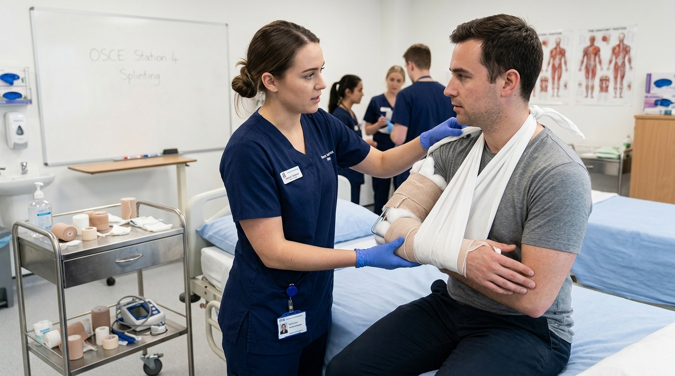
- Immobilize at least 2 adjacent joints (the joint above and the joint below the fracture site).
- Pre-mold the splint on the healthy limb of the patient or examiner before applying to the injured limb.
- Always pad bony prominences (olecranon, malleoli, fibular head) with cotton wool to prevent pressure ulcers, and assess distal pulses & capillary refill before and after application!
Hemostatic Tourniquet (Garou Hemostatic Esmarch) Protocol
Indications: Massive, arterial limb hemorrhage uncontrollable by direct wound compression or pressure dressing.
- Upper Extremity: Upper third of arm (sub-axillary). STRICTLY FORBIDDEN: Middle third of arm due to risk of radial nerve compression in the spiral groove (radial nerve palsy / "wrist drop")!
- Lower Extremity: Middle or upper third of thigh.
Rules of Tourniquet Application:
- Apply over a layer of cloth/clothing (never directly on bare skin to prevent skin necrosis).
- Tighten only until bleeding stops and distal pulse vanishes (avoid over-tightening).
- Attach a conspicuous written note: Exact time (hour and minute), date, and name of applicator.
- Maximum Duration: Summer: 1.5 – 2.0 hours; Winter: 45 – 60 minutes.
- Reperfusion Protocol: Every 30-45 minutes, loosen the tourniquet slowly for 3-5 minutes while holding manual digital pressure on the wound, then reapply slightly proximal.
✂️ 3. Surgical Instruments & Suturing Mastery
Tray Layout & Suture Types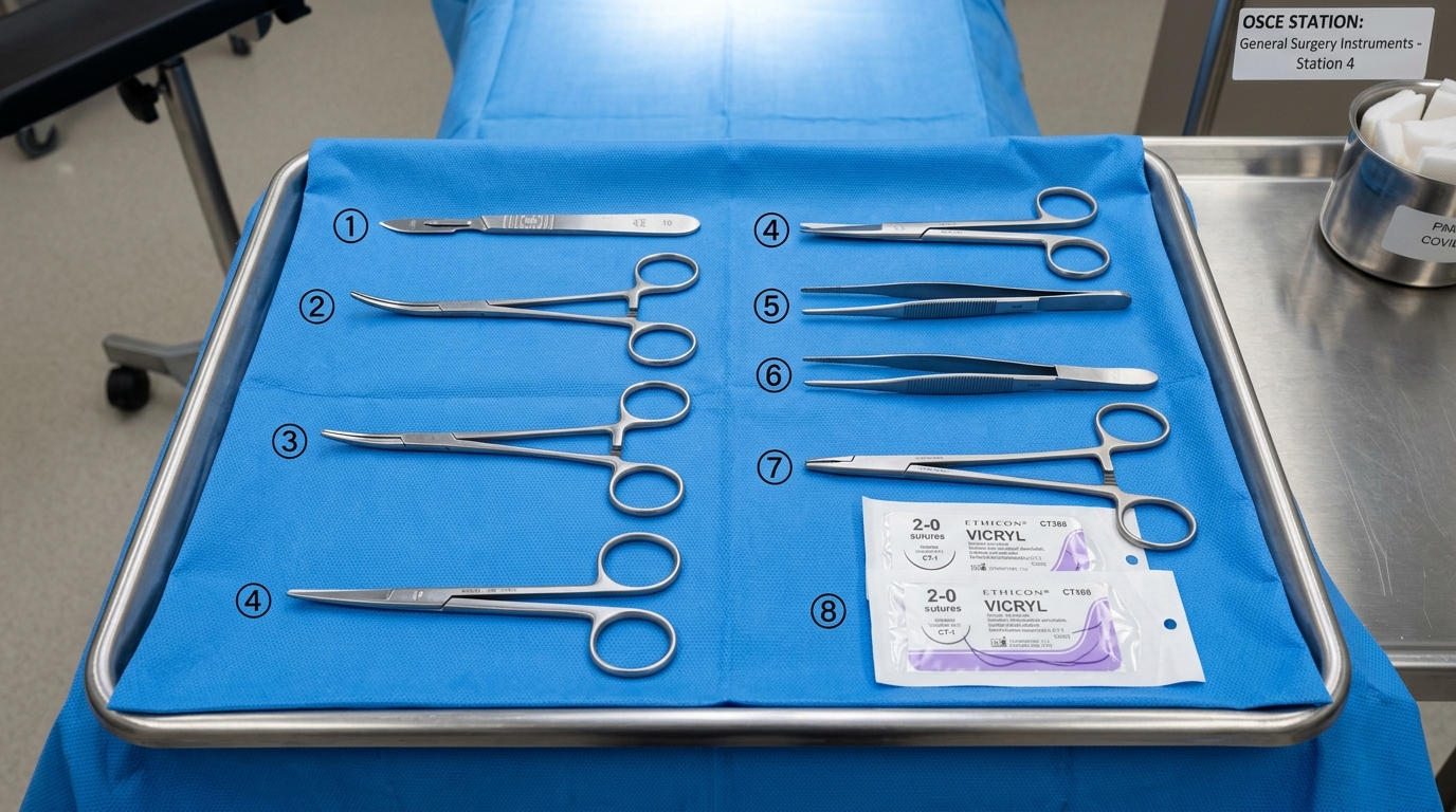
- Needle Holder (Hegar): Thumb in ring 1, ring finger in ring 2, index finger extended along shank for stabilization. Palmar grip used for heavy suturing.
- Tissue Forceps: Held like a pen (between thumb, index, and middle finger). Never hold like a dagger!
- Anatomical vs Surgical Forceps: Anatomical (smooth transverse grooves) is strictly used for delicate viscera (bowel, vessels, nerves). Surgical (toothed 1x2) is strictly for skin, fascia, and tough aponeurosis — NEVER on bowel or vessels!
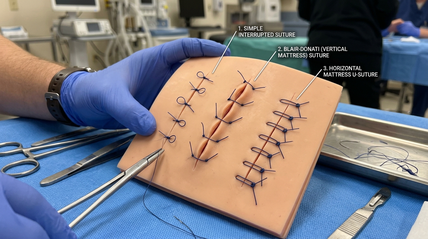
| Thread Type | Resorbable / Non-resorbable | Absorption Time | Best Surgical Indications |
|---|---|---|---|
| Catgut (Simple / Chromic) | Natural Resorbable (sheep intestine submucosa) | Simple: 7-10d Chromic: 21-28d |
Subcutaneous fat, rapid mucosal healing. High tissue reaction; rarely used today. |
| Vicryl (Polyglactin 910) | Synthetic Braided Resorbable | 56 – 70 days (tensile strength 50% at 21d) | Subcutaneous tissue, muscle, gastrointestinal anastomoses, biliary tract, urinary tract. |
| Monocryl / PDS | Synthetic Monofilament Resorbable | Monocryl: 90-120d PDS: 180-210d |
Subcuticular skin closure (Monocryl); slow-healing fascia, abdominal wall closure, pediatric surgery (PDS). |
| Silk (Mătase) | Natural Non-resorbable (protein braid) | Gradual degradation over 2 years | Vessel ligatures, bowel seromuscular sutures. Excellent handling and knot security. |
| Prolene (Polypropylene) | Synthetic Monofilament Non-resorbable | Permanent (no degradation) | Vascular anastomoses, hernia mesh fixation, abdominal fascia closure, cuticular skin. Zero tissue friction. |
| Nylon / Ethilon | Synthetic Monofilament Non-resorbable | Permanent / very slow hydrolysis | Standard skin suturing, tendon repair. High tensile strength, minimal inflammation. |
Clinical Manipulations, Tubes & Local Blocks
🩸 1. Blood Group Determination (ABO & Rh)
OSCE Essential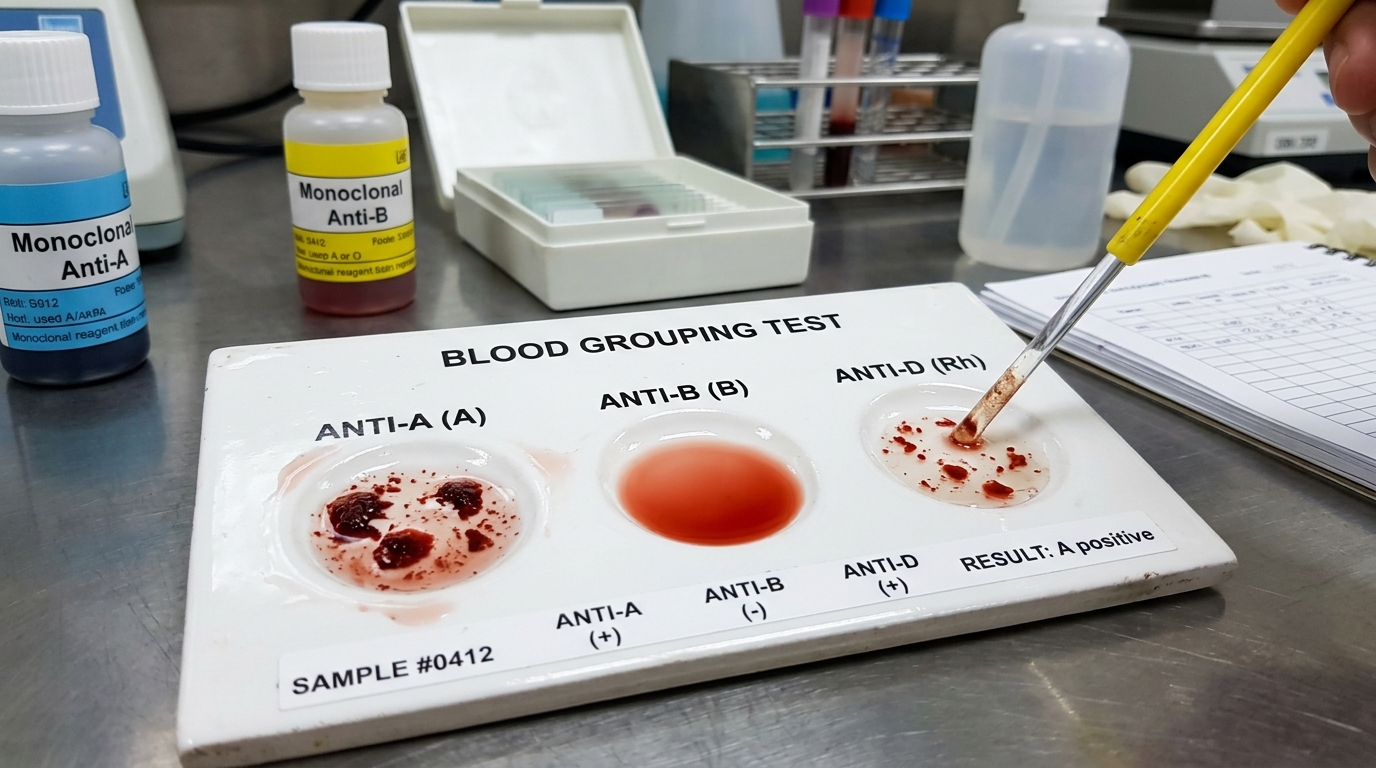
| Blood Group | Anti-A Reagent | Anti-B Reagent | Anti-AB Reagent | RBC Antigens & Serum Antibodies |
|---|---|---|---|---|
| Group O (I) | ❌ No agglutination | ❌ No agglutination | ❌ No agglutination | No A or B antigens; Anti-A and Anti-B antibodies |
| Group A (II) | ✅ Agglutination | ❌ No agglutination | ✅ Agglutination | Antigen A; Anti-B antibody |
| Group B (III) | ❌ No agglutination | ✅ Agglutination | ✅ Agglutination | Antigen B; Anti-A antibody |
| Group AB (IV) | ✅ Agglutination | ✅ Agglutination | ✅ Agglutination | Antigens A and B; No antibodies |
🫁 2. Sengstaken-Blakemore Tube & Kehr T-Tube
Special Surgical Tubes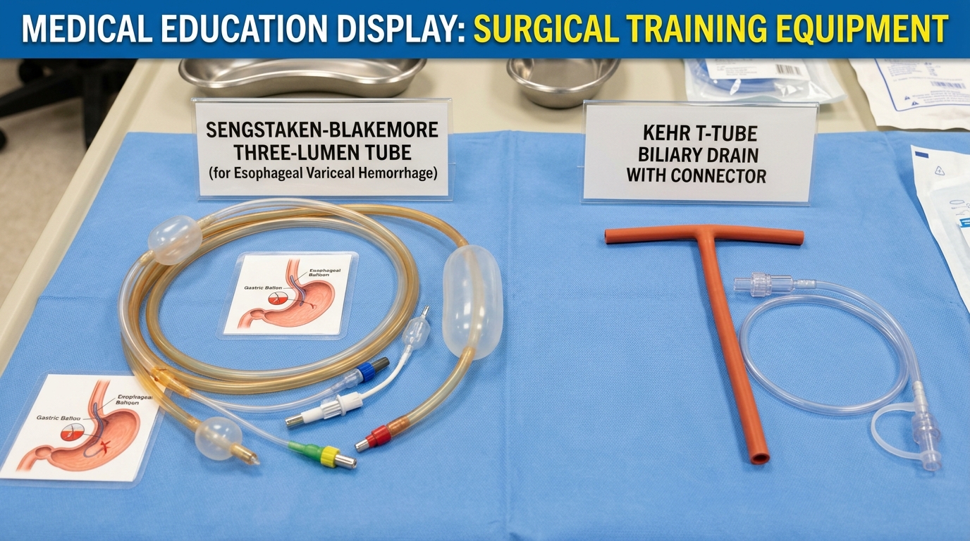
- Test balloons for leaks underwater; pass lubricated tube via nose into stomach.
- Inflate gastric balloon with 200–250 ml air, clamp port, pull snugly against cardia/GE junction, apply 0.5–1.0 kg traction.
- If bleeding continues: inflate esophageal balloon to 30–40 mmHg monitored with a manometer.
- Safety Rule: Deflate esophageal balloon every 12 hours for 15–30 minutes to prevent pressure necrosis and esophageal rupture! Max duration 24–48 hours.
- Placed in common bile duct (CBD) after choledochotomy / stone extraction.
- Timing: Kept in place for 12 to 14 days minimum to ensure a mature fibrous tract forms around the tube.
- Prerequisite 1: Clamping trial for 24–48 hours with NO abdominal pain, jaundice, fever, or leakage around the tube.
- Prerequisite 2: Fistulocholangiography (T-tube cholangiogram) showing free, rapid passage of contrast into the duodenum with normal CBD caliber and complete absence of residual calculi or strictures!
💉 3. Local Anesthetic Blocks, Catheters & Enemas
Manipulations ChecklistNovocaine Local Nerve Blocks
- Vishnevskii Block: "Creeping tight infiltration" (infiltrație târâtoare) using large volumes (200-500 ml) of dilute 0.25% procaine into fascial compartments for hydraulic dissection and deep regional block.
- Oberst-Lukashevich Block: Digital block for finger panaritium / injuries. Rubber band tourniquet at finger base, 1-2 ml 1% lidocaine on medial & lateral sides of proximal phalanx. CONTRAINDICATION: NEVER use adrenaline (causes digital gangrene!).
- Retromammary Block: Infiltrate 100-150 ml 0.25% novocaine behind mammary gland via submammary crease before incision of acute mastitis / abscess.
- Intercostal Block: Puncture upper border of rib below to reach lower border of upper rib; aspirate before injecting 3-5 ml 1% lidocaine.
Urinary Catheterization & NGT
- Foley Catheter: Instill 10 ml 2% lidocaine gel into male urethra; insert catheter until urine flows, advance extra 2-3 cm; inflate balloon with 5-10 ml sterile distilled water (never saline to avoid crystallization in valve!).
- Nasogastric Tube (NGT): Measure length: nose tip → earlobe → xiphoid process (~50-55 cm). Lubricate, advance as patient sips water. Verify position: auscultate epigastric whoosh with 20 ml air, aspirate acidic gastric juice (pH < 5.5).
- Post-Op Gastric Lavage: Intermittent infusion and siphoning of 300-500 ml warm saline in pyloric stenosis or gastric resection until returned aspirate is clear.
Enemas & Pulse Oximetry
- Cleansing Enema: 1.0–1.5 L warm water (36-37°C), left lateral Sims position, cannula inserted 8-10 cm, reservoir elevated 1 meter.
- Hypertonic Enema: 50–100 ml 10% NaCl or MgSO4; osmotic fluid draw stimulates strong peristalsis.
- Ognev Enema: 50 ml 10% NaCl + 30 ml 3% H2O2 + 50 ml Glycerin. Vigorous mechanical and osmotic stimulus for refractory paralytic ileus!
- Pulse Oximetry (SpO2): 660nm (red) and 940nm (infrared) spectrophotometry. Normal ≥ 95%. Pitfalls: hypoperfusion, nail polish, CO poisoning (falsely high), methemoglobinemia (~85%).
Special Surgical Pathology, Semiology & Signs
🩹 1. Acute Appendicitis: Points & Eponymous Signs
High Frequency- McBurney's Point: Junction of lateral 1/3 and medial 2/3 of spinoumbilical line (from right ASIS to umbilicus).
- Lanz's Point: Junction of right 1/3 and middle 1/3 of the bi-iliac line (between anterior superior iliac spines).
- Morris's Point: Medial 1/3 of the spinoumbilical line (closer to umbilicus).
- Sonnenburg's Point: Intersection of bi-iliac line with the lateral border of right rectus abdominis muscle.
| Clinical Sign | Examination Technique | Clinical & Pathological Significance |
|---|---|---|
| Dieulafoy Sign | Gentle stroking or pinching of skin over right iliac fossa (Sherren's triangle). | Cutaneous hyperesthesia indicating localized somatic peritoneal irritation. |
| Rovsing Sign | Pressure or percussion applied deeply in left iliac fossa. | Triggers sharp pain in right iliac fossa due to retrograde displacement of colonic gas distending the inflamed cecum. |
| Sitkovsky Sign | Patient is asked to roll from supine position onto the left side. | Exacerbation of right iliac fossa pain as the inflamed appendix and cecum drop and stretch inflamed mesentery. |
| Bartomier-Michelson | Palpation of right iliac fossa while the patient remains in the left lateral decubitus position. | Significantly more painful than supine palpation because abdominal muscles relax and cecum shifts forward. |
| Cope's Psoas Sign | Patient on left side; examiner passively hyperextends the right hip. | Pain indicates retrocecal appendix irritating the underlying iliopsoas muscle. |
| Cope's Obturator Sign | Patient supine; examiner flexes right hip and knee to 90° and internally rotates the thigh. | Pain in hypogastrium indicates pelvic appendix irritating internal obturator muscle. |
| Blumberg Sign | Slow, deep abdominal palpation followed by sudden, abrupt release of hand. | Sharp rebound tenderness — cardinal sign of peritoneal irritation (parietal peritonitis). |
| Mandel-Razdolsky Sign | Gentle fingertip percussion across abdominal quadrants. | Localized percussion pain over right iliac fossa — sign of localized peritonitis. |
🛡️ 2. Abdominal Wall Hernias: Differentiation
Inguinal & StrangulationInguinal canal, femoral canal (under inguinal ligament medial to femoral vein), umbilical ring, linea alba (epigastric hernias), linea semilunaris Spigelii, lumbar triangles of Petit (inferior) and Grynfeltt (superior).
- Sudden, acute, severe non-radiating pain at the hernia site.
- Tense, rock-hard, exquisitely tender, irreducible swelling.
- Total absence of expansile cough impulse (+ subsequent signs of mechanical bowel obstruction).
| Feature | Indirect (Oblique External) Inguinal Hernia | Direct (Internal) Inguinal Hernia |
|---|---|---|
| Anatomical Pathway | Enters deep ring lateral to inferior epigastric vessels; travels along inguinal canal within spermatic cord; exits external ring. | Bulges directly forward through Hesselbach's triangle medial to inferior epigastric vessels. |
| Deep Ring Occlusion Test | Finger over deep ring (midway between ASIS and pubic tubercle) PREVENTS hernia descent on coughing. | Hernia continues to bulge despite deep ring occlusion. |
| Scrotal Descent & Strangulation | Frequently descends into scrotum; narrow neck → High risk of strangulation. Common in youth. | Rarely reaches scrotum; wide neck → Low strangulation risk. Common in elderly men. |
| Post-Op Care | Application of a scrotal suspensory bandage post-herniotomy to eliminate dead space and prevent scrotal hematoma. | |
🩻 3. Intestinal Occlusion (Bowel Obstruction)
Radiology & Clinical Level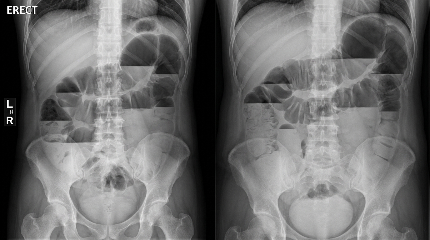
| Obstruction Level | Clinical Presentation | Radiological Distinctions (X-Ray) |
|---|---|---|
| High Small Bowel (Jejunal) | Early, intractable vomiting; severe dehydration; minimal abdominal meteorism; rapid metabolic alkalosis. | Few central fluid levels; valvulae conniventes (herringbone/feather pattern); no colon gas. |
| Low Small Bowel (Ileal) | Bilious to fecaloid vomiting; central step-ladder distension; colicky cramping pain; absolute obstipation. | Numerous Kloiber levels in center of abdomen; air-fluid levels are wider than tall. |
| Large Bowel (Colonic) | Massive peripheral distension along colonic frame; late fecaloid vomiting; obstipation. Closed-loop risk! | Peripheral air-fluid levels that are taller than wide; prominent haustrations (do not cross full lumen). |
💥 4. Peptic Ulcer: Perforation, Bleeding & Stenosis
Triad of Ulcer Complications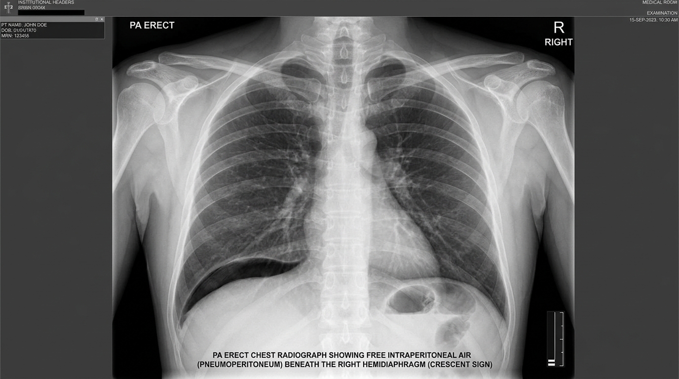
Sudden catastrophic "dagger-thrust" pain (Dieulafoy); board-like rigidity ("ventre de bois"); Jobert's sign (hepatic dullness replaced by percussion tympany); Spasokukotsky sign (complete peristaltic silence). If X-ray is inconclusive, perform pneumogastrography (inject 200-300 ml air via NGT) and repeat X-ray.
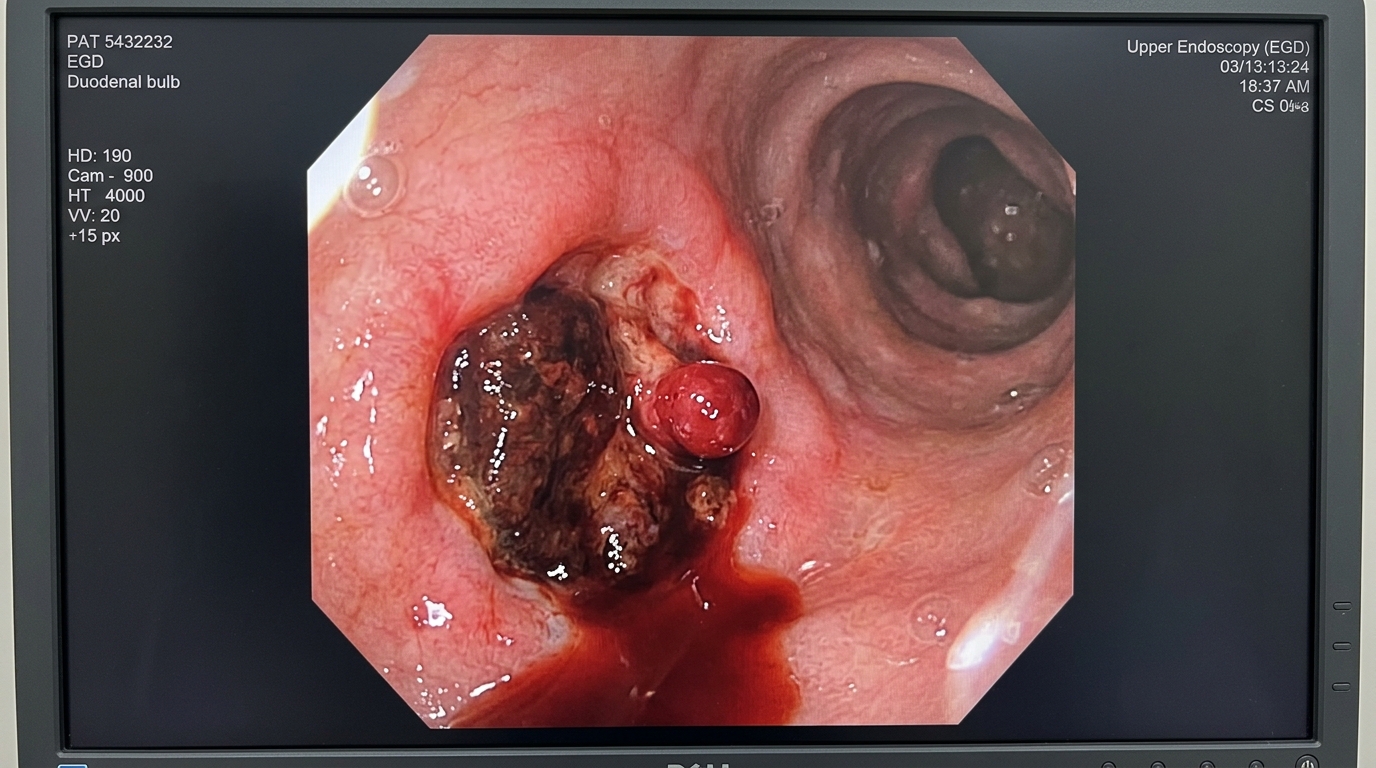
Forrest Class: Ia (Spurting arterial), Ib (Oozing), IIa (Non-bleeding visible vessel), IIb (Adherent clot), IIc (Black hematin base), III (Clean ulcer base).
Drug Protocol: IV Omeprazole 80 mg bolus → 8 mg/h infusion × 72h + Octreotide 50 mcg bolus → 50 mcg/h.
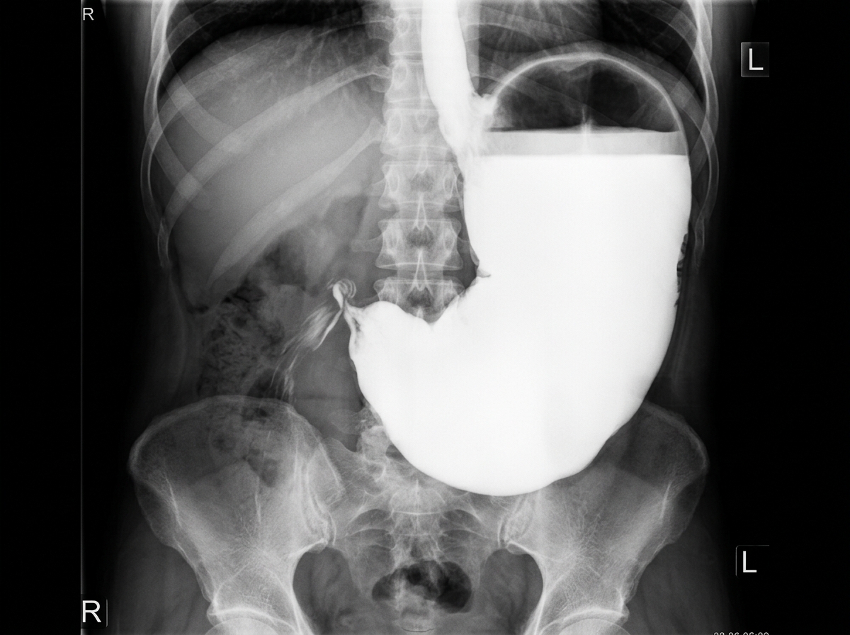
Patkin Triad: (1) Succussion splash ("clapotage"); (2) Visible peristalsis; (3) Asymmetric left hypochondrium fullness.
Kussmaul Sign: Visible peristaltic waves traveling left to right across epigastrium.
Darrow Syndrome: Severe persistent vomiting of HCl causes hypochloremic, hypokalemic metabolic alkalosis with paradoxical aciduria, secondary prerenal azotemia, and tetany. Barium meal shows 3 layers and >24h retention.
🟢 5. Gallbladder & Biliary Disease Semiology
Signs & Ultrasonography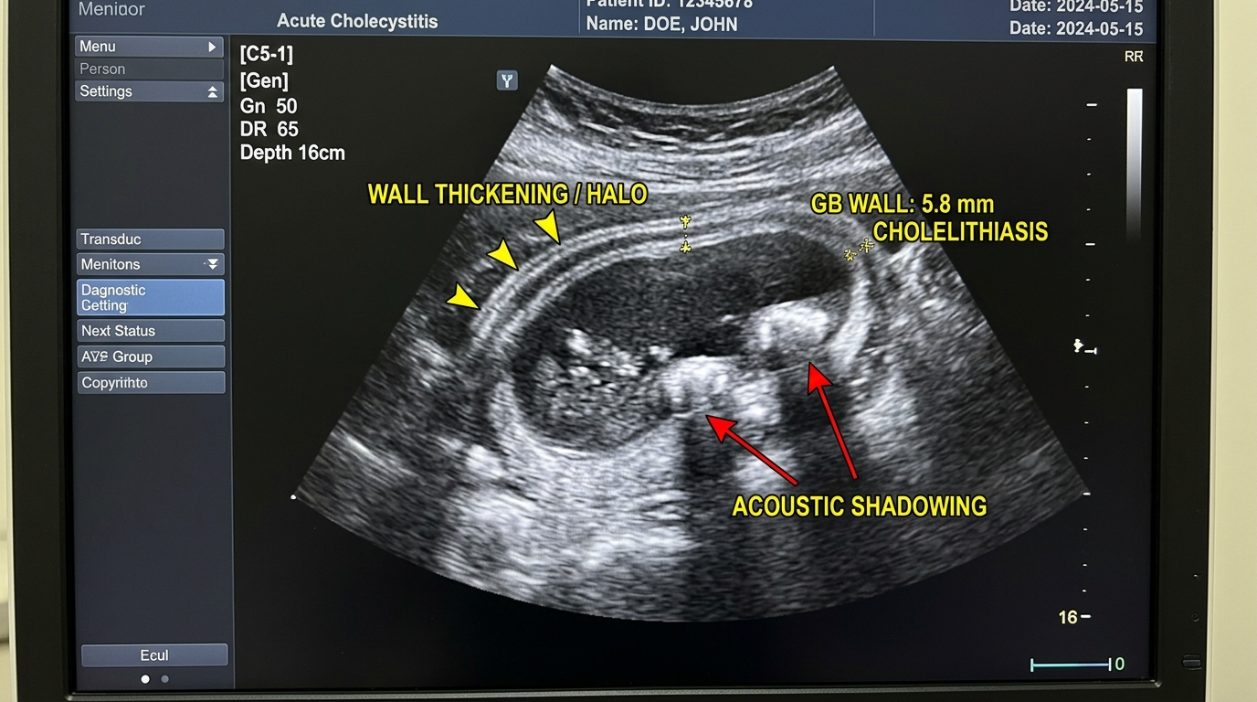
| Eponymous Sign | Execution & Findings | Clinical Significance |
|---|---|---|
| Murphy's Sign | Deep palpation under right costal margin while patient inhales deeply. | Sudden arrest of inspiration due to sharp acute pain when inflamed gallbladder contacts examiner's fingers. |
| Ortner's Sign | Gentle percussive tap with ulnar border of hand over right costal arch. | Sharp pain triggered over inflamed liver/gallbladder bed (Ortner's percussion symptom). |
| Mussie-Gheorghievsky Sign | Pressure applied between sternal and clavicular heads of right sternocleidomastoid muscle. | Exquisite tenderness — phrenic nerve sign due to diaphragmatic peritoneum irritation. |
| Courvoisier-Terrier Sign | Palpable, distended, non-tender gallbladder in a patient with progressive jaundice. | Suggests malignant periampullary or pancreatic head tumor obstruction (NOT gallstones). |
🔥 6. Acute Pancreatitis: Clinical Signs & CT
Severe Abdominal Emergency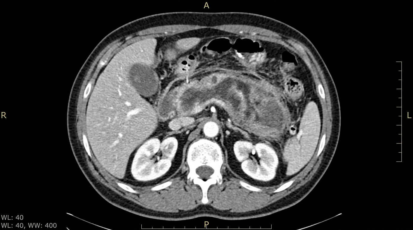
- Körte's Sign: Painful transverse band of muscular resistance 6-7 cm above umbilicus along the anatomical axis of the pancreas.
- Mayo-Robson's Sign: Exquisite tenderness on palpation in the left costovertebral angle (phrenico-costal angle).
- Voskresensky's Sign: Absence of palpable epigastric abdominal aortic pulsation due to pancreatic retroperitoneal inflammatory phlegmon.
- Grey Turner's Sign: Bluish-purple ecchymosis of the flanks (retroperitoneal dissection of hemorrhagic exudate).
- Cullen's Sign: Periumbilical ecchymosis (hemoperitoneum tracking along falciform ligament).
- Gobiet's Sign: Epigastric meteorism due to isolated transverse colon dilatation (colon cut-off sign).
🫁 7. Thoracic & Abdominal Trauma Emergencies
Life-Threatening Trauma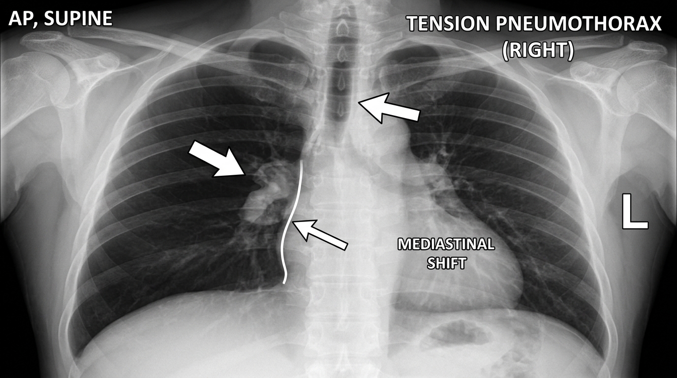
Severe dyspnea, tracheal deviation, absent breath sounds, hyperresonance, hypotension (obstructive shock). IMMEDIATE ACTION BEFORE X-RAY: Needle thoracocentesis with large-bore cannula (14-16G) in 2nd intercostal space midclavicular line (or 5th ICS AAL). Followed immediately by tube thoracostomy connected to underwater Bülau drain.
7th or 8th intercostal space on posterior axillary or scapular line. Puncture strictly at the UPPER BORDER OF THE LOWER RIB to avoid the intercostal neurovascular bundle (Vein-Artery-Nerve) located in the subcostal groove of the upper rib!
- Immediate initial drainage > 1,500 ml of blood upon chest tube insertion.
- Persistent bleeding > 200–300 ml/hour for 3–4 consecutive hours.
Aspiration of >10 ml gross blood, RBC count >100,000/mm³, WBC count >500/mm³, presence of bile, amylase, bacteria or food debris.
🩸 8. Venous Pathology & Varicose Vein Tests
DVT & Functional Tests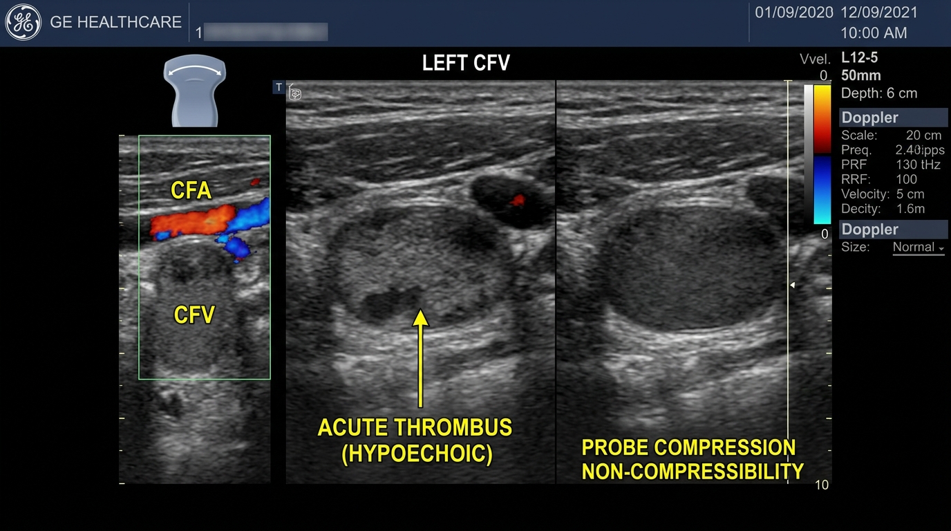
| Functional Test | Method & Execution | Pathological Interpretation |
|---|---|---|
| Brodie-Trendelenburg-Troyanov | Patient supine, leg elevated to empty veins; apply tourniquet over saphenofemoral junction; patient stands. | Rapid filling from above upon releasing tourniquet = Saphenofemoral junction incompetence. Rapid filling within 30s before release = Incompetent perforator veins. |
| Perthes Test | Tourniquet placed below knee on standing patient (veins distended); patient walks for 5-10 minutes. | Veins collapse = Deep venous system is patent. Veins remain distended/painful = Deep venous obstruction (Absolute contraindication to stripping!). |
| Delbet Test | Tourniquet applied around upper thigh; patient walks. | Evaluates functional patency of the muscular calf veno-motor pump. |
| Pratt I & II Tests | Double elastic bandage applied from toes upwards; unwrap lower bandage on standing patient. | Locates exact anatomical site of incompetent perforator veins where veins bulge between wraps. |
| Sheinis Test | Three tourniquets applied: upper thigh, above knee, below knee. | Segmentally isolates saphenofemoral, saphenopopliteal, and perforator vein incompetence. |
| Homans & Moses Signs | Homans: calf pain on forced dorsiflexion of foot. Moses: calf pain on anteroposterior compression against tibia. | Cardinal physical signs of Deep Vein Thrombosis (DVT). |
Paraclinical Investigation Master Reference
📊 Complete Blood Count, Chemistry & Coagulation Master Table
Surgical Interpretation Guide| Investigation | Standard Normal Range | Elevated In Surgical Pathology | Decreased In Surgical Pathology | Surgical Significance & Red Flags |
|---|---|---|---|---|
| Hemoglobin (Hb) | Male: 130–170 g/L Female: 120–150 g/L |
Hemoconcentration (severe pancreatitis, bowel obstruction, burns, dehydration) | Acute gastrointestinal bleeding, ruptured spleen/liver, chronic malnutrition/malignancy | Grade I anemia (90-110), Grade II (70-90), Grade III (<70 g/L). Transfusion threshold usually <70-80 g/L. |
| Hematocrit (Hct) | Male: 40–52% Female: 36–48% |
Severe dehydration, hypovolemia, hemoconcentration (>44% in pancreatitis indicates severe necrosis) | Active hemorrhage (after fluid resuscitation), chronic blood loss | Reliable gauge of intravascular fluid depletion and hemoconcentration. |
| WBC & Band Forms | WBC: 4.0–9.0 × 10⁹/L Bands: 1–5% |
Appendicitis, peritonitis, phlegmon, acute cholecystitis, bowel strangulation | Severe sepsis / septic shock (depleted marrow), viral infections | Left shift (Bands > 6-10%): Hallmarked in gangrenous appendicitis, purulent peritonitis, infected necrosis! |
| Total & Direct Bilirubin | Total: 8.5–20.5 μmol/L Direct: 0–5.1 μmol/L |
Direct (conjugated) >50%: Choledocholithiasis, periampullary cancer, biliary stricture (mechanical jaundice). Indirect >80%: Hemolysis. | N/A | Dark urine and clay-colored pale stools characteristic of mechanical/obstructive jaundice. |
| ALT, AST & ALP, GGT | ALT: 7–56 U/L ALP: 44–147 U/L GGT: 9–48 U/L |
ALP & GGT elevated >3x: Obstructive biliary pathology / choledocholithiasis. AST/ALT high: Hepatocellular necrosis. |
N/A | Differential: High ALP/GGT + High Direct Bilirubin = Biliary obstruction! |
| Serum Amylase & Lipase | Amylase: 28–100 U/L Lipase: 10–140 U/L |
Acute pancreatitis (>3x upper limit), perforated duodenal ulcer (moderate rise), bowel infarction | End-stage pancreatic destruction, chronic fibrosis | Lipase is significantly more specific than amylase and remains elevated for 8–14 days. |
| Serum Calcium (Ca²⁺) | 2.15–2.55 mmol/L (Total) 1.15–1.33 mmol/L (Ionized) |
Hyperparathyroidism, bone metastasis | Severe acute pancreatitis (<2.0 mmol/L), Darrow syndrome | Hypocalcemia in pancreatitis is due to enzymatic fat necrosis and saponification; cardinal prognostic marker! |
| Electrolytes (Na⁺, K⁺, Cl⁻) | Na⁺: 135–145 mmol/L K⁺: 3.5–5.0 mmol/L Cl⁻: 96–106 mmol/L |
Hyperkalemia in crush syndrome, acute renal failure, massive hemolysis | Darrow Syndrome (Pyloric Stenosis): Hypochloremia (<90), Hypokalemia (<3.5), causing metabolic alkalosis! | Hypokalemia causes cardiac arrhythmias, paralytic ileus, and muscle weakness. Supplement IV with 0.9% saline! |
| Arterial Blood Gas & Lactate | pH: 7.35–7.45 HCO3⁻: 22–26 mmol/L Lactate: 0.5–2.0 mmol/L |
Lactate > 2.0–4.0 mmol/L in bowel ischemia/gangrene, hemorrhagic shock, severe septic peritonitis | Low pH and low HCO3⁻ indicate metabolic acidosis | Serial lactate clearance is the best clinical gauge of shock resuscitation adequacy. |
| Coagulogram (PT/INR, aPTT, D-Dimer) | INR: 0.8–1.2 aPTT: 25–35s D-Dimer: <500 ng/mL |
D-Dimer > 500–5000 in acute DVT and pulmonary embolism; prolonged INR/aPTT in liver failure, DIC, or warfarin | Fibrinogen < 1.5 g/L in consumptive coagulopathy / DIC | Prerequisite before any surgery: INR ≤ 1.5, Platelets ≥ 50–100 × 10⁹/L. |
Standardized Patient Diagnostic Simulator (12 Clinical Cases)
📊 Paraclinical Laboratory Profile:
| WBC | 17.8 × 10⁹/L (Normal: 4.0–9.0) | Leukocytosis with Left Shift |
| Band Neutrophils | 18% (Normal: 1–5%) | Marked inflammatory shift |
| CRP | 88 mg/L (Normal: <5.0) | High acute-phase reactant |
| Urinalysis | Clear, occasional RBCs (3-4/hpf) | Ureteral proximity irritation |
🩻 Ultrasound Findings:
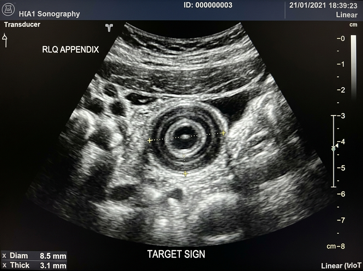
Non-compressible, blind-ending tubular structure in the right iliac fossa measuring 9.2 mm in outer diameter (>6 mm). "Target sign" on cross-section with wall hypervascularity on color Doppler. Local periappendiceal fat stranding and trace free fluid.
✅ Complete Diagnostic Formulation:
Primary Diagnosis: Acute phlegmonous-gangrenous appendicitis with localized peritonitis.
Paraclinical Justification: Marked leukocytosis (17.8 × 10⁹/L) with significant left shift (18% bands) and high CRP (88 mg/L) confirming bacterial transmural inflammation. Ultrasound reveals non-compressible appendix >9 mm with target sign and surrounding free fluid.
Differential Diagnoses: Acute mesenteric lymphadenitis (common in youth, multiple enlarged lymph nodes), Meckel's diverticulitis, acute terminal ileitis (Crohn's), right renal colic (severe radiating loin-to-groin pain, prominent hematuria), gynecological pathology in females (ruptured ovarian cyst, ectopic pregnancy).
Preoperative & Emergency Plan: NPO, IV fluid resuscitation (crystalloids), IV analgesia (avoid masking peritoneal signs until surgeon reviews), prophylactic single-dose broad-spectrum antibiotics (Ceftriaxone 2g IV + Metronidazole 500mg IV), urgent laparoscopic or open appendectomy within 12–24 hours.
📊 Paraclinical Laboratory Profile:
| WBC | 16.2 × 10⁹/L (Normal: 4.0–9.0) | Severe leukocytosis |
| Serum Amylase | 260 U/L (Normal: 28–100) | Moderate reactive rise |
| ABG pH | 7.31 • Lactate: 3.2 mmol/L | Metabolic lactic acidosis |
| Hb / Hct | 145 g/L / 43% | Baseline normal / hemoconcentration |
🩻 Erect Chest Radiograph:
✅ Complete Diagnostic Formulation:
Primary Diagnosis: Perforated duodenal (or prepyloric) peptic ulcer complicated by acute generalized chemical-purulent peritonitis; pneumoperitoneum.
Paraclinical Justification: Erect chest X-ray demonstrates the pathognomonic subdiaphragmatic gas sickle (free intra-abdominal air confirming hollow visceral perforation). Moderate hyperamylasemia is typical due to peritoneal absorption of gastroduodenal fluid. High lactate (3.2 mmol/L) reflects systemic hypoperfusion and peritoneal inflammation.
Differential Diagnoses: Acute pancreatitis (hyperamylasemia >3x normal, epigastric pain radiating to back, absence of subdiaphragmatic free air on X-ray), acute cholecystitis with perforation, acute myocardial infarction (inferior wall), high small bowel strangulation.
Preoperative & Emergency Management: Immediate NPO; insert wide-bore NGT on continuous active suction to decompress stomach and prevent further peritoneal leakage; vigorous IV crystalloid resuscitation (2–3 L Ringer's lactate); IV high-dose PPI (Omeprazole 80 mg IV bolus); broad-spectrum IV antibiotics covering Gram-negatives and anaerobes (Ceftriaxone + Metronidazole); emergency laparotomy/laparoscopy: Graham patch omentopexy closure of perforation, copious peritoneal lavage, and abdominal cavity drainage.
📊 Paraclinical Laboratory Profile:
| Serum Lipase | 3,450 U/L (Normal: 10–140) | >24x elevation (diagnostic) |
| Serum Amylase | 1,280 U/L (Normal: 28–100) | >12x elevation |
| Serum Calcium | 1.72 mmol/L (Normal: 2.15–2.55) | Critical Hypocalcemia |
| Hematocrit (Hct) | 48.5% (Normal: 40–52%) | Severe hemoconcentration |
| CRP (48h) | 280 mg/L (Normal: <5) | Necrotizing marker (>150) |
🩻 Abdominal CT Contrast:
✅ Complete Diagnostic Formulation:
Primary Diagnosis: Severe acute necrotizing pancreatitis (Balthazar grade E, CT severity index 8/10), complicated by retroperitoneal inflammatory phlegmon, hypocalcemia, and systemic inflammatory response syndrome (SIRS).
Paraclinical Justification: Serum lipase elevated >24x normal; CT scan demonstrates diffuse pancreatic parenchymal necrosis and extensive peripancreatic collections. Critical hypocalcemia (1.72 mmol/L) is a direct consequence of enzymatic fat necrosis (pancreatic lipases break down retroperitoneal triglycerides into free fatty acids which saponify and bind ionized calcium into insoluble soaps). Elevated Hct (48.5%) reflects massive capillary leak into retroperitoneal third space.
Differential Diagnoses: Perforated peptic ulcer (has free air on X-ray, sudden dagger onset), acute mesenteric ischemia (severe pain out of proportion to exam, high lactate), acute cholecystitis, aortic dissection.
Intensive Management Protocol: Admission to ICU; aggressive targeted fluid resuscitation with Ringer's lactate (250–500 ml/h titrated to urine output ≥0.5–1.0 ml/kg/h and Hct normalization); multi-modal analgesia (epidural analgesia or opioids with spasmolytics); IV Calcium gluconate supplementation; strict NPO in hyperacute phase; early enteral nutrition via nasojejunal tube once hemodynamics stabilize; Somatostatin/Octreotide; prophylactic antibiotics NOT routinely recommended unless infected necrosis is proven by CT-guided FNA.
📊 Paraclinical Laboratory Profile:
| Potassium (K⁺) | 3.1 mmol/L (Normal: 3.5–5.0) | Hypokalemia (loss in vomitus) |
| Chloride (Cl⁻) | 89 mmol/L (Normal: 96–106) | Hypochloremia |
| Urea / Creatinine | 14.8 mmol/L / 130 μmol/L | Prerenal azotemia (dehydration) |
| Lactate | 1.8 mmol/L (Normal: <2.0) | No strangulation / ischemia yet |
🩻 Erect Abdominal X-Ray:
✅ Complete Diagnostic Formulation:
Primary Diagnosis: Acute mechanical small bowel obstruction (adhesive ileus secondary to pelvic postoperative adhesions); dehydration and secondary prerenal azotemia.
Paraclinical Justification: Plain erect abdominal radiograph shows multiple central step-ladder air-fluid levels (Kloiber cups) with plicae circulares and collapse of the colon, characteristic of mechanical small bowel occlusion. Electrolytes demonstrate hypochloremic hypokalemic dehydration with elevated BUN/creatinine ratio (prerenal azotemia). Normal lactate indicates absence of bowel strangulation/gangrene at present.
Differential Diagnoses: Paralytic (adynamic) ileus (diffuse gas in both small bowel and colon with sluggish/absent bowel sounds), strangulated groin hernia (always examine femoral and inguinal orifices!), large bowel obstruction (colonic dilatation >6cm, haustrations), acute mesenteric ischemia.
Preoperative & Emergency Protocol: Nasogastric decompression with Salem sump tube on continuous suction; vigorous fluid resuscitation with 0.9% NaCl + KCl supplementation (correct hypokalemia to prevent cardiac arrhythmia); catheterize bladder (hourly diuresis monitoring); perform water-soluble contrast follow-through (Gastrografin / Schwartz test) to evaluate transit; if no contrast enters cecum by 24h or if peritoneal signs, fever, leukocytosis or elevated lactate develop → immediate exploratory laparotomy, adhesiolysis, and bowel viability assessment.
📊 Paraclinical Laboratory Profile:
| Chloride (Cl⁻) | 76 mmol/L (Normal: 96–106) | Severe Hypochloremia |
| Potassium (K⁺) | 2.6 mmol/L (Normal: 3.5–5.0) | Severe Hypokalemia |
| Serum Sodium (Na⁺) | 128 mmol/L (Normal: 135–145) | Hyponatremia |
| Arterial Blood Gas | pH: 7.56 • HCO3⁻: 38 mmol/L | Severe Metabolic Alkalosis |
| Urine pH | 5.2 (Paradoxical Aciduria) | Pathognomonic of Darrow Syn. |
| Urea / Creatinine | 18.5 mmol/L / 180 μmol/L | Prerenal azotemia |
🩻 Barium Meal Examination:
Enormous gastromegaly: the greater curvature of the stomach sags deeply into the true pelvic cavity. The pyloric canal is narrowed to a thread-like lumen with complete absence of duodenal bulb filling. Three-layer phenomenon: dense barium residue at bottom, layer of clear fluid in middle, and air/food debris at the top. Retention of >80% of barium in stomach after 24 hours.
✅ Complete Diagnostic Formulation:
Primary Diagnosis: Cicatricial pyloroduodenal stenosis in the decompensated stage secondary to chronic peptic ulcer disease, complicated by Darrow syndrome (hypochloremic, hypokalemic metabolic alkalosis with paradoxical aciduria, hypocalcemic tetany, dehydration, and secondary prerenal azotemia).
Pathophysiological Mechanism of Darrow Syndrome: Massive loss of gastric hydrochloric acid (H⁺ and Cl⁻) leads to primary metabolic alkalosis. In response to hypovolemia, aldosterone acts on the distal renal tubule to reabsorb Na⁺, but because Cl⁻ is depleted, the kidney must excrete K⁺ and H⁺ in exchange for Na⁺, producing paradoxical aciduria in the face of systemic alkalosis! Severe hypokalemia and secondary hypocalcemia (alkalosis increases calcium binding to albumin) trigger muscle tetany and carpopedal spasm.
Differential Diagnoses: Antral gastric adenocarcinoma (older age, anorexia, cachexia, endoscopy shows irregular ulcerated mass), gastroparesis (diabetic, no mechanical obstruction on barium/endoscopy), duodenal compression by superior mesenteric artery (Wilkie's syndrome).
Preoperative & Emergency Management: Daily saline gastric lavage via large-bore Levin tube (clears food debris, reduces mucosal edema and bacterial fermentation); aggressive correction of metabolic alkalosis with IV 0.9% Normal Saline + extensive KCl infusion (30–40 mEq/L at ≤20 mEq/h; never administer glucose solutions without potassium as glucose drives K⁺ into cells and worsens paralysis); IV calcium gluconate for tetany; total parenteral nutrition (TPN); elective definitive surgery once metabolic panel normalizes (Truncal Vagotomy + Gastrojejunostomy, or Antrectomy + Billroth I/II).
📊 Paraclinical Laboratory Profile:
| Hemoglobin (Hb) | 68 g/L (Normal: 130–170) | Severe Acute Anemia (Grade III) |
| Hematocrit (Hct) | 21.5% (Normal: 40–52%) | Critical hemodilution |
| RBC Count | 2.2 × 10¹²/L | Marked normocytic anemia |
| BUN / Urea | 16.4 mmol/L | GI blood protein absorption |
| Serum Creatinine | 92 μmol/L (Normal) | Normal baseline renal status |
| Coagulogram | INR: 1.1 • aPTT: 28s | No underlying coagulopathy |
🩻 Urgent Upper Endoscopy (EGD):
✅ Complete Diagnostic Formulation:
Primary Diagnosis: Acute upper gastrointestinal hemorrhage from a chronic duodenal bulb ulcer (posterior wall); Forrest Class IIa/Ib (visible non-bleeding vessel with active oozing); severe acute post-hemorrhagic anemia (Grade III); Class III hypovolemic-hemorrhagic shock (Algöwer Shock Index 1.47).
Paraclinical Justification: Critical Hb of 68 g/L and Hct 21.5% in the presence of tachycardia and hypotension indicate acute blood loss >30–40% of circulating blood volume (>1,500 ml). Disproportionate elevation of blood urea nitrogen (BUN) with normal creatinine is pathognomonic of upper GI bleeding, resulting from massive breakdown and absorption of digested red blood cell proteins in the small intestine.
Differential Diagnoses: Bleeding esophageal/gastric varices (history of cirrhosis, stigmata of portal hypertension), Mallory-Weiss tear (violent vomiting preceding hematemesis), Dieulafoy lesion, hemorrhagic gastritis, gastric cancer.
Emergency Management Protocol: Two large-bore peripheral IV lines (16G or 14G); immediate crystalloid infusion followed by matched Packed Red Blood Cells (PRBCs) with target Hb 70–90 g/L; IV Omeprazole 80 mg bolus followed by 8 mg/h continuous infusion for 72 hours; Tranexamic acid 1g IV; urgent therapeutic EGD: dual endoscopic hemostasis (epinephrine injection 1:10,000 + bipolar electrocoagulation or hemoclip placement); if endoscopic hemostasis fails → urgent transcatheter arterial embolization (TAE) or emergency laparotomy with duodenotomy and transfixion ligation of the gastroduodenal artery (GDA).
📊 Paraclinical Laboratory Profile:
| Total Bilirubin | 118 μmol/L (Normal: 8.5–20.5) | Marked hyperbilirubinemia |
| Direct Bilirubin | 94 μmol/L (Normal: 0–5.1) | >80% conjugated (mechanical) |
| Alkaline Phos. (ALP) | 485 U/L (Normal: 44–147) | Cholestasis marker (>3x) |
| GGT | 320 U/L (Normal: 9–48) | Marked biliary obstruction |
| ALT / AST | 85 U/L / 72 U/L | Mild secondary hepatocyte injury |
| WBC | 15.4 × 10⁹/L (Bands 12%) | Bacterial infection |
🩻 Abdominal Ultrasonography (USG):
✅ Complete Diagnostic Formulation:
Primary Diagnosis: Acute phlegmonous calculous cholecystitis complicated by choledocholithiasis (secondary gallstone migration into CBD), mechanical (obstructive) jaundice, and acute cholangitis (Charcot's Triad: RUQ pain + Jaundice + Fever).
Paraclinical Justification: Biochemical profile demonstrates prominent cholestasis: conjugated (direct) bilirubin constitutes >80% of total bilirubin, accompanied by a >3x elevation of ALP and GGT, with acholic stools and bilirubinuria. Ultrasound confirms acute cholecystitis criteria (thickened gallbladder wall 5.8 mm, pericholecystic halo, calculi with acoustic shadows) and common bile duct dilation (11 mm > 6-7 mm) with intraductal calculus.
Differential Diagnoses: Periampullary / Pancreatic head adenocarcinoma (Courvoisier-Terrier sign: painless palpable gallbladder + progressive jaundice, whereas in choledocholithiasis pain is prominent and gallbladder is non-distensible due to chronic fibrosis), acute viral hepatitis (AST/ALT >1000 U/L, minimal ALP rise), hemolytic jaundice (indirect bilirubin >80%, dark stools, normal ALP/GGT).
Preoperative & Emergency Management: NPO, IV fluid resuscitation, analgesics (Drotaverine, Papaverine; avoid morphine); broad-spectrum IV antibiotics covering Gram-negatives and anaerobes (Ceftriaxone 2g IV + Metronidazole 500mg IV q8h); urgent ERCP (CPGRE) with endoscopic sphincterotomy and balloon/basket stone extraction to decompress the infected biliary tree; followed by laparoscopic cholecystectomy during the same hospital admission.
📊 Paraclinical Laboratory Profile:
| Hemoglobin (Hb) | 82 g/L (down from baseline) | Acute internal hemorrhage |
| Hematocrit (Hct) | 25% (Normal: 40–52%) | Acute blood loss |
| Arterial Blood Gas | pH: 7.28 • Lactate: 4.1 mmol/L | Hypoperfusion lactic acidosis |
| Base Deficit | -7.5 mmol/L | Class III hemorrhagic shock |
🩻 Diagnostic Imaging & FAST Exam:
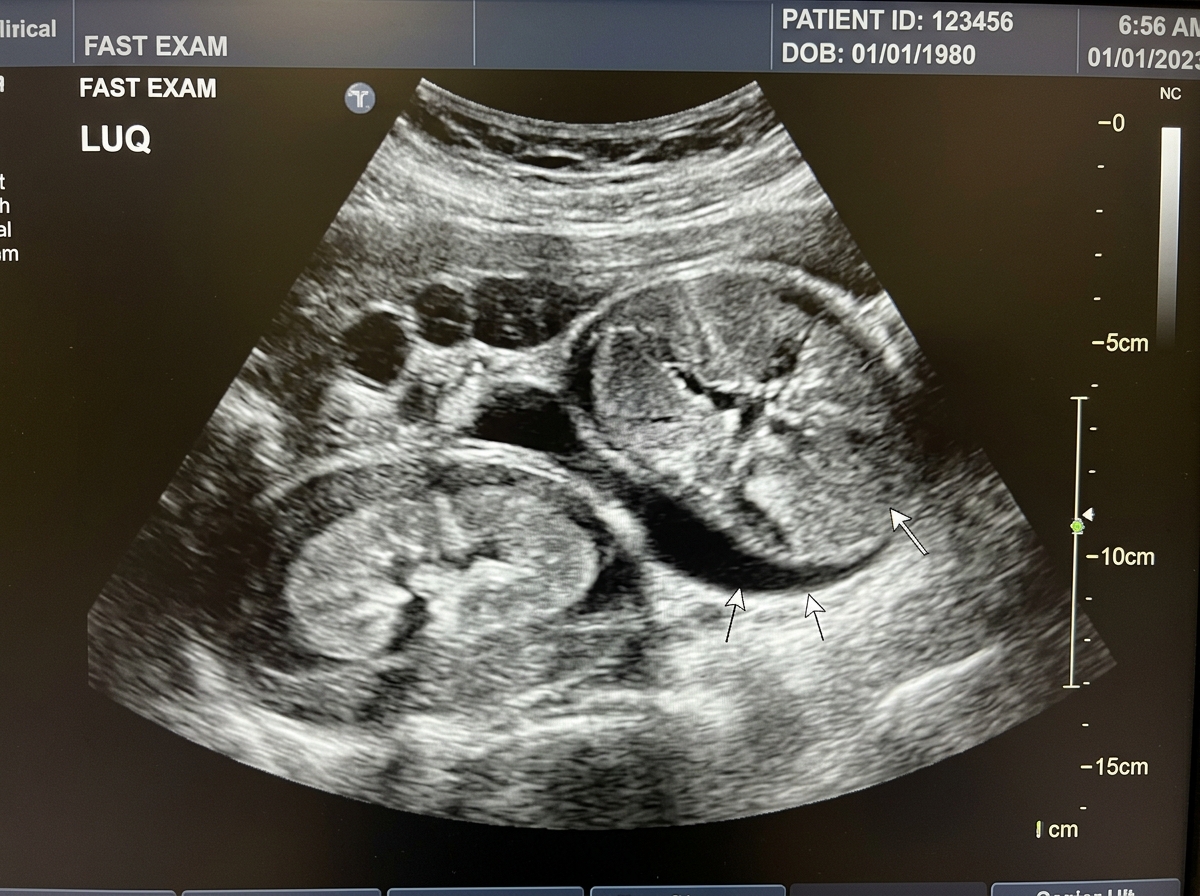
FAST Ultrasound (Bedside): Positive for free fluid in Morison's pouch (hepatorenal space), splenorenal recess, and retrovesical space (Pouch of Douglas). Disruption of spleen lower pole parenchyma with subcapsular hematoma.
Diagnostic Peritoneal Lavage (Laparocentesis): Aspiration of 15 ml gross, non-clotting dark blood immediately on catheter insertion (strongly positive criteria >10 ml blood).
✅ Complete Diagnostic Formulation:
Primary Diagnosis: Closed blunt abdominal trauma; Grade IV splenic rupture with massive hemoperitoneum; left 9th-10th rib fractures; Class III hemorrhagic-hypovolemic shock.
Paraclinical Justification: Kehr's sign (referred left shoulder pain mediated via the C3-C5 phrenic nerve due to blood and clots irritating the undersurface of the left hemidiaphragm). Bedside FAST ultrasound and DPL confirm massive hemoperitoneum. High lactate (4.1 mmol/L) and base deficit (-7.5 mmol/L) demonstrate acute tissue hypoperfusion.
Differential Diagnoses: Liver laceration (RUQ tenderness, Morison's pouch hematoma), retroperitoneal hematoma secondary to pelvic fracture, mesenteric avulsion, hollow viscous rupture (would show free air on X-ray, peritonitis).
Emergency Surgical Plan: Immediate massive transfusion protocol (1:1:1 ratio of PRBCs, Fresh Frozen Plasma, and Platelets); permissive hypotension (target SBP 80-90 mmHg until surgical hemostasis); immediate transfer to the operating theater for emergency exploratory laparotomy, splenectomy (or splenorrhaphy if viable in minor tear), evacuation of hemoperitoneum, and inspection of all four abdominal quadrants.
📊 Paraclinical Profile & ABG:
| PaO2 | 48 mmHg (Normal: 80–100) | Life-threatening hypoxia |
| PaCO2 | 54 mmHg (Normal: 35–45) | Acute hypercapnia / hypoventilation |
| pH | 7.22 | Mixed respiratory & lactic acidosis |
| Lactate | 4.8 mmol/L | Obstructive shock tissue hypoxia |
🩻 Chest Radiograph (Simulated Emergency):
✅ Complete Diagnostic Formulation:
Primary Diagnosis: Closed thoracic trauma; right-sided tension (suffocating/valvular) pneumothorax; total atelectasis of right lung; acute obstructive cardiogenic shock; acute respiratory failure.
Pathophysiological Mechanism: A one-way flap valve injury to the lung parenchyma or bronchus permits air to enter the pleural space during inspiration but prevents it from escaping during expiration. Intrapleural pressure progressively exceeds atmospheric pressure, completely collapsing the ipsilateral lung, depressing the diaphragm, and shifting the mediastinum, trachea, and heart to the contralateral side. This mechanical compression kinks the inferior and superior vena cava, obliterating venous return to the right heart, culminating in catastrophic obstructive shock and cardiac arrest!
Critical Examiner Rule: Tension pneumothorax is a STRICTLY CLINICAL DIAGNOSIS. NEVER DELAY TREATMENT TO OBTAIN A RADIOGRAPH! If the exam prompt states signs of tension pneumothorax, state immediately: "I will decompress the chest immediately before sending for an X-ray!"
Immediate Step-by-Step Life-Saving Protocol:
- Immediate Needle Decompression: Insert a large-bore cannula (14–16G, ≥5 cm length) into the 2nd intercostal space in the midclavicular line on the right side, directly above the 3rd rib. (Alternative per ATLS 10th ed: 4th/5th ICS anterior axillary line). A sudden hiss of escaping air confirms successful decompression and converts tension into simple pneumothorax.
- Definitive Tube Thoracostomy (Chest Drain): Insert a 28–32 Fr chest tube in the 5th intercostal space anterior to the mid-axillary line, directed posteriorly and superiorly, connected to an underwater seal drainage system (Bülau drain).
📊 Paraclinical Laboratory Profile:
| D-Dimer | 4,850 ng/mL (Normal: <500) | Marked active fibrinolysis |
| Platelet Count | 245 × 10⁹/L | Normal baseline |
| PT / INR | 13.2s / 1.05 | Normal coagulation profile |
| Serum Creatinine | 88 μmol/L | Normal renal function for LMWH |
🩻 Vascular Duplex Ultrasound:
✅ Complete Diagnostic Formulation:
Primary Diagnosis: Acute deep vein thrombosis (DVT) of the right iliofemoral venous system; high risk of pulmonary embolism (PE); Caprini thromboembolic score: High Risk (7 points).
Paraclinical Justification: Marked elevation of D-dimer (>4,800 ng/mL) demonstrates extensive ongoing endovascular fibrin clot turnover. Duplex Doppler ultrasound provides definitive confirmation by demonstrating the pathognomonic triad: (1) Incomplete or absent compressibility of the vein under transducer pressure, (2) Direct visualization of intraluminal thrombus, and (3) Absence of spontaneous color Doppler flow and loss of respiratory phasicity.
Differential Diagnoses: Ruptured Baker's cyst (synovial cyst of popliteal fossa, "pseudo-thrombophlebitis"), acute cellulitis/erysipelas (intense erythema with clear raised borders, high fever, normal D-dimer), superficial thrombophlebitis (palpable superficial tender indurated cord along great saphenous vein), gastrocnemius muscle tear/hematoma.
Therapeutic & Prophylactic Management:
- Immediate therapeutic anticoagulation: Low Molecular Weight Heparin (LMWH - Enoxaparin 1 mg/kg SC every 12 hours) or Direct Oral Anticoagulant (DOAC: Rivaroxaban 15 mg BID × 3 weeks → 20 mg daily, or Apixaban 10 mg BID × 7d → 5 mg BID).
- Limb elevation above heart level to facilitate venous return; bed rest initially until effective anticoagulation, followed by gradual mobilization with graduated class II elastic compression stockings.
- If anticoagulation is strictly contraindicated (e.g. active major intracranial/GI bleed) → deploy temporary Inferior Vena Cava (IVC) filter to prevent fatal pulmonary embolism.
📊 Paraclinical Profile:
| Procalcitonin | 24.5 ng/mL (Normal: <0.5) | Severe bacterial septic shock |
| WBC Count | 21.5 × 10⁹/L (Bands 24%) | Toxic shift-to-left |
| Lactate | 4.6 mmol/L | Severe tissue hypoperfusion |
| Peritoneal Fluid | Foul-smelling turbid purulent-fecal | Free bacterial contamination |
🩻 Abdominal CT Findings:
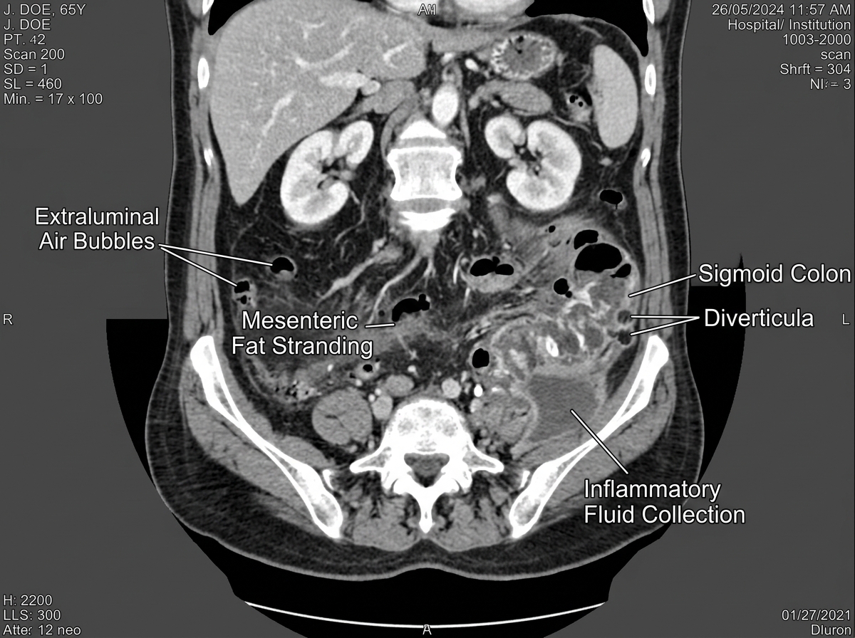
Thick-walled sigmoid colon with extensive diverticula and focal defect; massive pneumoperitoneum with free air in Morrison's pouch and inter-loop spaces; abundant dense free fluid in the pelvis and paracolic gutters (fecal peritonitis - Hinchey Stage IV).
✅ Complete Diagnostic Formulation:
Primary Diagnosis: Acute perforated sigmoid diverticulitis (Hinchey Stage IV); acute generalized purulent-fecal peritonitis; severe septic shock; toxic paralytic ileus.
Management: Sepsis bundle resuscitation: broad-spectrum antibiotics (Piperacillin-Tazobactam or Meropenem), IV crystalloids + vasopressors (Norepinephrine), emergency laparotomy: Hartmann's Procedure (sigmoid resection, end colostomy, rectal stump closure) with copious peritoneal toilet and multiple quadrant drains.
📊 Paraclinical Profile:
| Blood Glucose | 17.2 mmol/L (Normal: 3.9–6.1) | Severe diabetic decompensation |
| WBC Count | 18.9 × 10⁹/L | Marked neutrophilic leukocytosis |
| ESR | 65 mm/h | High systemic inflammation |
| Urine Ketones | Positive (+) | Ketoacidosis risk |
🔬 Clinical Features & Terminology:
Note on Medical Terminology: In Eastern European / Moldovan surgical literature, a surgical Carbuncle is traditionally termed "Cărbune / Anthrax chirurgical" (derived from French anthrax, distinct from Bacillus anthracis zoonosis). It represents confluent necrotizing staphylococcal furunculosis of multiple adjacent hair follicles extending into the deep subcutaneous fascia.
✅ Complete Diagnostic Formulation:
Primary Diagnosis: Carbuncle ("Anthrax chirurgical") of the posterior cervical region (nape); Type 2 Diabetes Mellitus in decompensated ketoacidotic state; severe soft tissue phlegmonous infection.
Management: Aggressive sliding-scale IV insulin therapy to normalize blood glucose; broad-spectrum anti-staphylococcal IV antibiotics (Oxacillin / Cefazolin or Vancomycin); urgent surgical intervention: wide cruciate / X-shaped or H-shaped incision, radical excision of all necrotic subcutaneous tissue down to the muscular fascia, hydrogen peroxide and chlorhexidine irrigation, and loose packing with hypertonic saline / povidone-iodine gauze.
Rapid-Fire Eponymous Signs Flashcard Deck
Murphy's Sign
Deep palpation under right costal margin while patient breathes in deeply.
Result:Sudden arrest of inspiration due to sharp pain when inflamed gallbladder contacts examiner's fingers. Pathognomonic for Acute Cholecystitis.
Rovsing's Sign
Deep palpation or percussion in the left iliac fossa.
Result:Elicits pain in the right iliac fossa due to retrograde displacement of gas through the colon distending the inflamed cecum and appendix.
Blumberg's Sign
Gradual deep palpation of the abdominal wall followed by sudden, abrupt release of hand pressure.
Result:Severe sharp rebound pain indicating parietal peritoneal irritation / Peritonitis.
Jobert's Sign
Percussion over the right anterior midclavicular line along the hepatic area.
Result:Disappearance of normal liver dullness, replaced by tympanic resonance due to free subdiaphragmatic gas (pneumoperitoneum from perforated ulcer).
Patkin's Triad
- Gastric succussion splash ("clapotage") heard >4-6h after fasting.
- Visible gastric peristalsis through abdominal wall.
- Asymmetric left upper quadrant gastric distension.
Kussmaul's Sign
Visible, forceful peristaltic waves rippling from the left hypochondrium across the epigastrium toward the right, seen through a thin, emaciated abdominal wall in decompensated pyloric stenosis.
Körte's Sign
A painful, transverse band of muscular resistance and tenderness across the epigastrium, 6–7 cm above the umbilicus along the anatomical axis of the pancreas in Acute Pancreatitis.
Mayo-Robson Sign
Exquisite tenderness elicited by palpation or pressure in the left costovertebral angle (phrenico-costal angle) corresponding to the inflamed tail of the pancreas.
Voskresensky's Sign
Inability to palpate the abdominal aortic pulse in the epigastrium due to retroperitoneal edema and inflammatory phlegmon surrounding the pancreas in Acute Pancreatitis.
Grey Turner & Cullen
Grey Turner: Blue-purple discoloration of the flanks (retroperitoneal blood).
Cullen: Blue-purple discoloration periumbilical (hemoperitoneum tracking via falciform ligament).
Kehr's Sign
Severe acute pain at the tip of the left shoulder exacerbated by lying supine. Caused by blood/clots from a ruptured spleen irritating the left hemidiaphragmatic peritoneum (phrenic nerve C3–C5).
Homans & Moses Signs
Homans: Calf pain upon passive forced dorsiflexion of the foot.
Moses: Calf pain elicited on anteroposterior compression against tibia (lateral compression painless). Diagnostic of DVT.
Live OSCE Section Scorer & Examiner Rubric
📝 5-Section Live Score Tracker
Bandage application, Kramer splint, tourniquet protocol, simple/Blair-Donati sutures.
Blood group ABO/Rh typing, Foley catheter, local blocks, NGT, Sengstaken & Kehr tube care.
X-rays (pneumoperitoneum, Kloiber levels), CT (pancreatitis), USG (cholecystitis), Endoscopy (Forrest).
CBC left-shift, Darrow syndrome, enzymes (lipase, amylase), hypocalcemia, ABG lactate.
Physical exam signs (Murphy, Rovsing, Blumberg), complete surgical diagnosis, pre-op plan.
Grading scale: 9–10 (Grade 10, Outstanding); 7–8 (Grade 8–9, Distinction); 5–6 (Grade 5–6, Minimum Pass); <5 (Unattested).